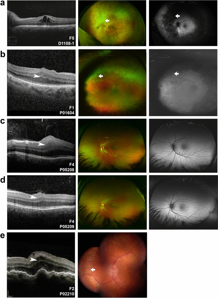
Figure 4.
Clinical imaging features of MFRP families. (a–e) OCT (left), Optos wide field photographs (a–d, middle) or composite fundus photograph (e, middle), and Optos fundus autofluorescence imaging of patients with biallelic MFRP variants (a–d, right). Three patients (a,b,e) had evidence of retinal degeneration in a characteristic ring pattern of atrophy (arrows). Two siblings (c,d) had very similar clinical appearance with macular folds (arrowheads), foveal hypoplasia, punctate hyperautofluorescent white lesions, crowded discs and vascular tortuosity. P02210 had prominent choroidal folds and foveoschisis (e).

