Figure 5.
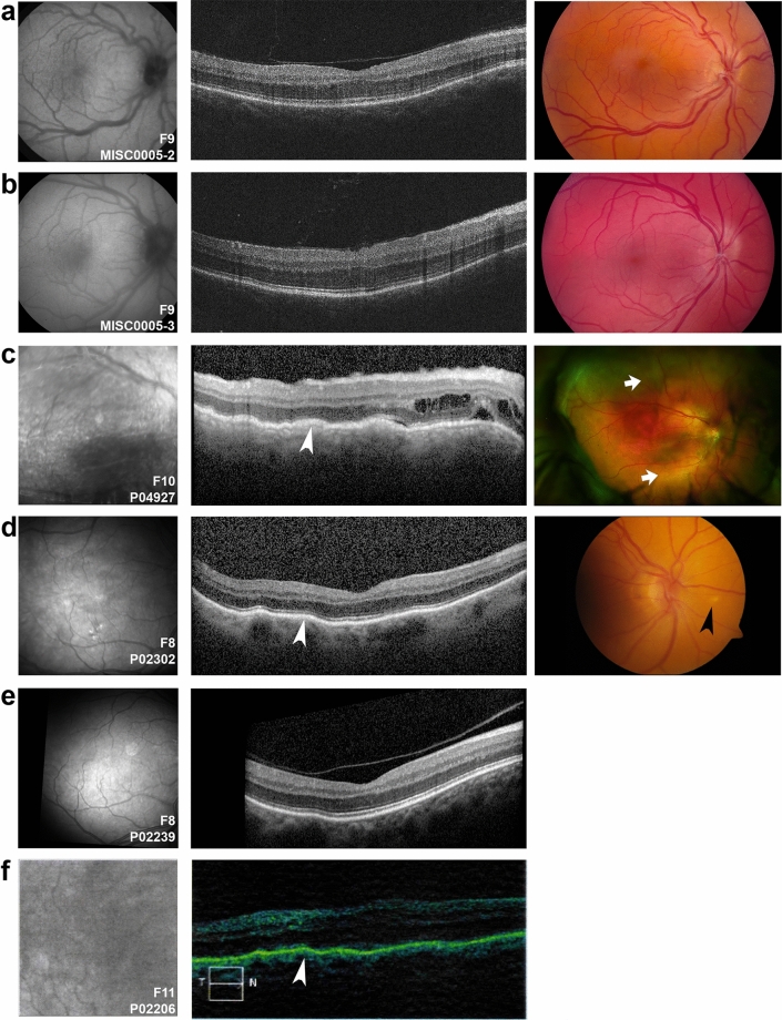
Clinical imaging features of PRSS56 families. (a,b) TopCon fundus autofluorescence (left) and color (right) and OCT (middle) images showing mild hypoautofluorescence in the macula, crowded discs, vascular tortuosity, and foveal hypoplasia among two family members carrying PRSS56 deleterious variants (MISC0005-2, F10, A) and (MISC0005-3, F10, B). (c–f) Infrared reflectance (left) and corresponding OCT image (middle) of patients carrying PRSS56 biallelic variants, along with Optos wide-field imaging (c, right) or optic disc image (d, right). P04927 (F11) has a serous retinal detachment along with intraretinal fluid (a, arrow) and pigmentary changes. Siblings P02302 and P02239 from F9 have similar clinical appearance with choroidal folds (white arrowheads) and mild foveal hypoplasia, with P02302 having white lesions in the retina (black arrowhead). These patients have prominent choroidal folds (c–f).

