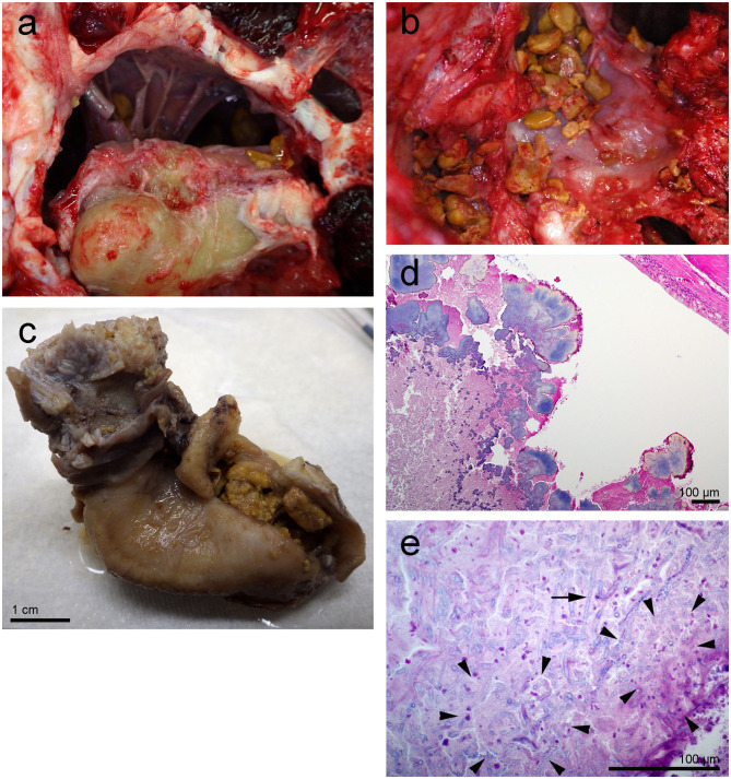
Figure 2.
Left tympanic bone from the beluga Qila. The gross images, (a,b) were taken during the necropsy and dissection of the ears. Note the smooth nodular tan yellow precipitates in the peribullar (a,b) and tympanic cavities (c). Image (b) was obtained after the removal of the tympano-periotic complex. (d,e) Histopathology of hematoxylin-eosin stained left tympanic bone and cavity with numerous lobules of bacteria (d) and florid fungal hyphae interspersed [arrow in (e)] within inflammatory infiltrate and necrotic debris [regions delimited by arrowheads in (e)].

