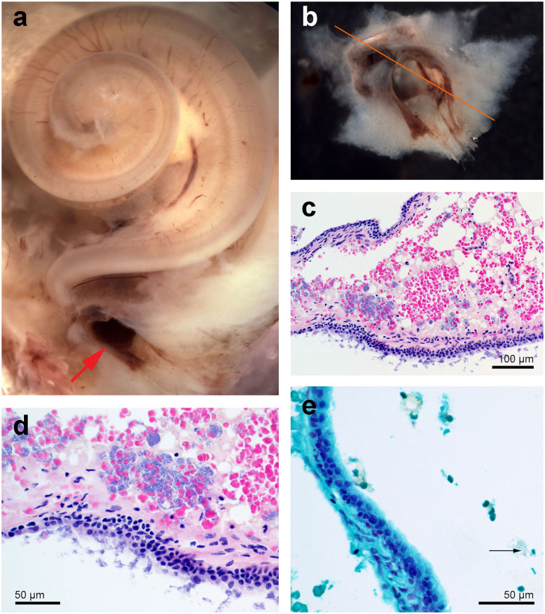
Figure 5.
Right cochlea from the beluga Qila. (a) General picture. Note there is multifocal congestion with hemorrhage at the round window (red arrow). (b) Detail of the round window niche after being sampled for histopathology. (c,d) Histopathology of focally extensive acute hemorrhage admixed with vacuolated proteinaceous deposits and dense clusters of extracellular (c,d) and rare intracellular [arrow in (e)] Gram positive cocci. Sections (c,d) were stained with hematoxylin-eosin and (e) with Twort's gram stain.

