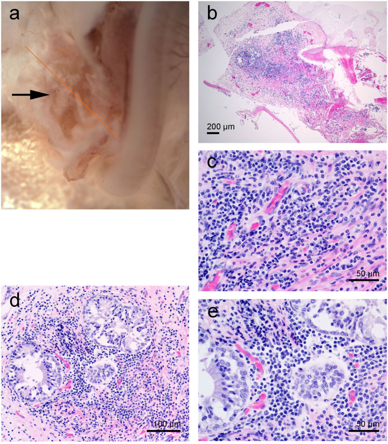
Figure 6.
Left cochlea from the beluga Aurora. (a) Sub-gross image of the base of the cochlea. The arrow highlights the white deposits at the round window niche. (b–d) Micrographs of Hematoxylin and Eosin stained sections with moderate focal accumulation of lymphocytes, plasma cells and fewer macrophages with scattered neutrophils. In images (d,e) there are a small number of entrapped variably sized glands.

