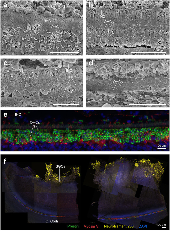
Figure 7.
(a–d) Scanning electron micrographs of the left cochlea from the beluga Aurora. Organ of Corti features three rows of outer hair cells (OHCs) and one row of inner hair cells (IHCs) in the upper (a) and lower (b) apical turn, upper (c) and lower (d) basal turn. (e,f) Right cochlea from the beluga Aurora. Immunofluorescence images of the middle turn (e) and hook or most basal part of the right cochlea. (f) The beginning of the hook is located at the left the image, which is the region of the cochlea where the highest frequencies are encoded. Flat preparations of the cochlea were incubated with anti- prestin (green), anti-myosin VI (red), anti-neurofilament 200 kD (yellow) antibodies and DAPI (blue). SGCs, spiral ganglion cells.

