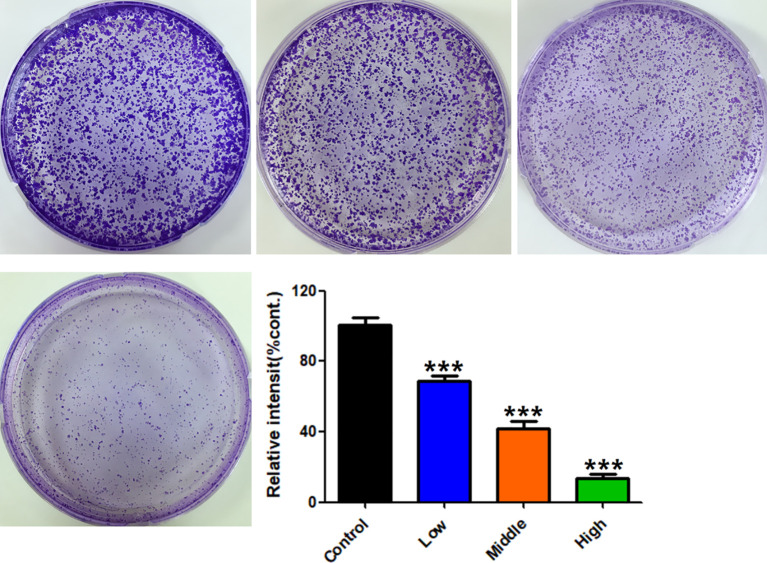
Figure 8.
Effects of RPL on cloning formation of A549 cells. Cells were treated with RPL (0.2, 0.4, and 0.6 mg/mL) for 14 days and then stained with crystal violet. The colony formation was observed under a microscope, and five fields were randomly selected for counting. Data were expressed as mean ± SD (n = 3), and asterisk indicated significant difference, ***p < 0.001 vs. control.

