Figure 5.
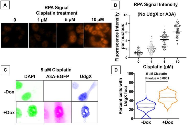
Cisplatin treatment increases cellular RPA and UdgX foci. (A) HEK293T cells were treated with cisplatin at the different concentrations and RPA was visualized using antibodies. (B) The RPA signal intensity of cells in part A was quantified at each concentration. (C) HTO-A3A-EGFP cells were induced for A3A-EGFP expression using doxycycline and then treated with cisplatin and transfected with pFLAG-UdgX. The fluorescence image colors are inverted for clarity. (D) The cells from part C were used to quantify the number of cells with UdgX foci. The median values are shown using dashed lines (- - - -), while the upper and lower quartiles are shown using light lines (___).

