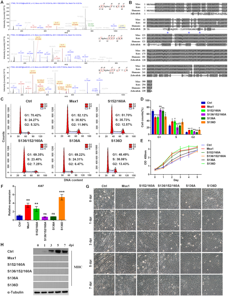
Figure 4.
Msx1 promotes C2C12 cell proliferation via the phosphorylation of Ser136. (A) Tandem mass spectrometry of the Msx1 peptides modified by phosphorylation on the Ser136 (upper), Ser152 (middle) and Ser160 (lower) residues. (B) Alignment of Msx1 proteins across different species in chordates. The asterisks indicate Ser136, Ser152 and Ser160 respectively. (C) Flow cytometry assays to analyze the cell cycle of C2C12 cells overexpressing different Msx1 mutants. (D) Statistical analysis of the cell cycle of C2C12 cells overexpressing different Msx1 mutants. Values are the means ± SD. **P < 0.001, *P < 0.01. (E) CCK-8 assays to examine the proliferation of C2C12 cells overexpressing different Msx1 mutants. Values are the means ± SD. (F) qRT-PCR assays to detect Ki67 mRNA levels in C2C12 cells overexpressing different Msx1 mutants. Values are the means ± SD. ***P < 0.0001, **P < 0.001. (G) Micrographs of C2C12 cells at 0, 1, 3, 5 and 7 dpi with overexpression of different Msx1 mutants to assess the impact of Msx1 phosphorylation sites on the inhibition of differentiation. (H) Western blotting to detect the marker of terminal muscle differentiation MHC. C2C12 cells overexpressing different Msx1 mutants were harvested and lysed separately at 0, 1, 3, 5 and 7 dpi. Western blotting assays were performed to detect MHC expression.

