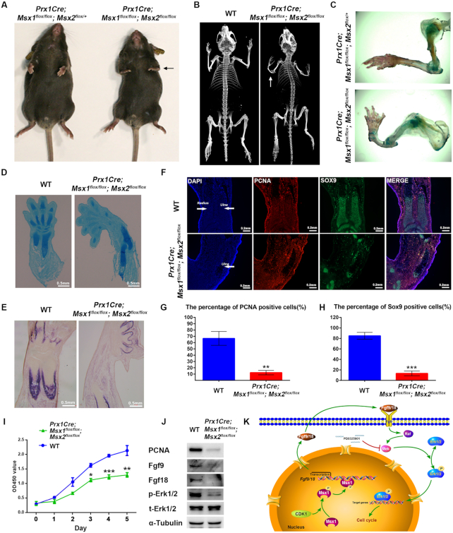
Figure 7.
Deficiency of Msx in the lateral plate mesoderm reduced bone formation. (A) Representative views of 4-week-old control and Msx1/2 MSC-specific knockout littermates. The black arrow indicates the forelimb. (B) μCT images of bones from 4-week-old wild-type and Msx1/2 MSC-specific knockout mice. (C) Alcian blue and ARS staining in the forelimbs from 4-week-old control and Msx1/2 MSC-specific knockout mice. (D, E) Alcian blue (D) and ALP (E) staining of limb sections from wild-type and Msx1/2 MSC-specific knockout mice at postnatal day 2 (P2), scale bar = 0.5 mm. (F) IF assays of limb sections from wild-type and Msx1/2 MSC-specific knockout mice at P2. Expression of Sox9 (green) and PCNA (red) was detected. DAPI was used to counterstain the nuclei. Scale bar = 0.2 mm. (G, H) Diagram summarizing the immunofluorescence data. The proportions of PCNA (G)- and Sox9 (H)-positive cells (n = 400) were calculated. Values are the means ± SD. ***P < 0.0001, **P < 0.001. (I) CCK-8 assays to evaluate the proliferation of primary bone marrow MSCs from wild-type and Msx1/2 MSC-specific knockout mice. Values are the means ± SD. ***P < 0.0001, **P < 0.001, *P < 0.01. (J) Western blotting to detect PCNA, p-Erk1/2, Fgf9 and Fgf18 levels in primary bone marrow MSCs from wild-type and Msx1/2 MSC-specific knockout mice. (K) Model of the promotion of cellular proliferation by Msx1. In the nucleus, Ser136 of Msx1 is phosphorylated by CDK1. Phosphorylated Msx1 binds to Fgf9 and Fgf18 to activate their transcription. Upregulated Fgf9 and Fgf18 are shuttled to the extracellular matrix, where they bind to Fgfrs to activate the MAPK signaling pathway and promote the phosphorylation of Erk1/2. p-Erk1/2 then promotes cell proliferation in a variety of ways.

