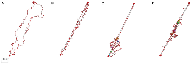
Figure 2.
Geometry of simulated plasmid chromosome. 3D geometry of simulated polymer model after reaching equilibrium. Red beads are the discretized units connected through springs (black segments). Two red boxes represent centromeres that are fixed in space. (A) Geometry of the raw polymer chain model. (B) Geometry of the polymer model with histones. Loops along the strand show locations of histones. (C) Geometry of the polymer model with condensins. Large spheres with the same color are the attachment sites of a condensin complex. (D) Geometry of the polymer model with both histones and condensins.

