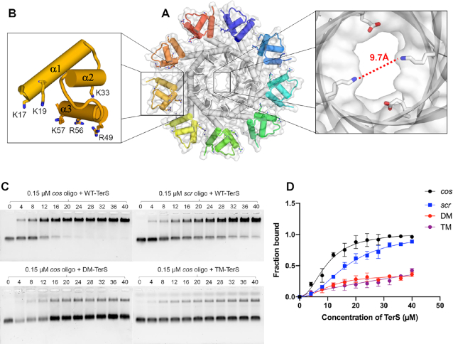
Figure 6.
DNA-binding activity of PaP3 TerS. (A) Solvent surface view of PaP3 TerS with a magnified view of the central channel, which has a minimum diameter of 9.7 Å, too narrow to accommodate dsDNA. (B) Zoom-in view of the N-terminal HTH domain; basic residues, putatively involved in DNA-binding, are shown as sticks. (C) Native agarose gel electrophoresis of PaP3 WT-TerS binding to Cy3-cos (top left) and Cy3-scr DNA (top right) as well as DM-TerS (bottom left) and TM-TerS (bottom right) binding to Cy3-cos. In all gels, a fixed concentration of cos DNA was titrated against 0–40 μM of TerS. (D) Quantification of band shift data in panel C. The error bar represents the standard deviation calculated from three independent gels.

