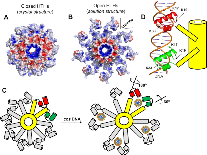
Figure 7.
Proposed model for TerS recognition of packaging initiation sites by DNA-interdigitation. (A) Electrostatic surface potential of TerS with closed HTHs observed in the crystal structure and (B) the open conformation of TerS modeled based on the solution structure. In both panels, the electrostatic surface potentials range between −5 kT/e representing negative charges (colored in red) and +5 kT/e representing positive charges (colored in blue). (C) Schematic top-view of PaP3 TerS: the oligomerization core and two nearby protomers are colored in yellow while the reminder seven protomers are in gray. The HTH helices α1–α2 are colored in green (protomer #1) and red (protomer #2). Four ds-DNA molecules are modeled as interdigitated by four pairs of TerS protomers. (D) Schematic representation of a 20-mer DNA oligonucleotide laterally interdigitated by two subunits of TerS whose HTH helixes α1 and α2 are color-coded as in panel (C).

