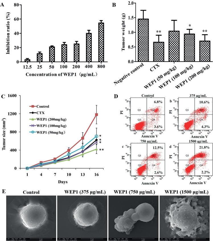
Fig. 2.
Effects of WEP1 on the growth inhibition of H22 cells and H22-bearing mice. (A) Inhibitory effect in vitro of WEP1 against H22 cells at 24 h treatment. (B, C) Inhibitory effect of polysaccharide WEP1 on H22 tumor growth in BALB/c mice. *P < 0.05 and **P < 0.01 compared with negative control group. (D) WEP1-induced apoptosis in H22 cells. Apoptosis was assayed by flow cytometry after cells were treated without (a, control) or with (b) 375, (c) 750, and (d) 1,500 μg/mL of WEP1 for 24 h. (E) Scanning electron micrographs (magnification ×6,000) of H22 cells showed the surface morphology changes of the cells. Data were presented as means ± SD of three independent experiments. Values were based on eight mice in each group.

