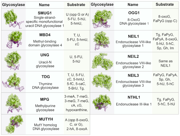
Figure 3.
BER glycosylases, their structures and respective substrates. The glycosylases (green) are bound to DNA (purple) containing a lesion (purple, space-filled). All structures are human except SMUG1 (Xenopus laevis), MUTYH (Geobacillus stearothermophilus), NEIL2 (Monodelphis domestica), NEIL3 (Mus musculus), NTHL1 (EndoIII, Geobacillus stearothermophilus). PDB: SMUG1 (1OE4), MBD4 (5CHZ), UNG (1EMH), TDG (3UFJ), MPG (1BNK), MUTYH (4YOQ), OGG1 (1EBM), NEIL1 (5ITY), NEIL2 (6VJI), NEIL3 (3W0F), NTHL1 (1ORN). Abbreviations: U, uracil; A, adenine; T, thymine; C, cytosine; G, guanine; 5-FU, 5-fluorouracil; 5-hmU, 5-hydroxymethyluracil; ϵ, etheno; FaPy, 2,6-diamino-4-hydroxy-5-N-methylformamidopyrimidine; 8-oxoG, 8-oxoguanine; Gh, Guanidonohydantoin; Sp, Spiroiminodihydantonin; Im, iminoallantoin; 5fC, 5-formylcytosine; 5caC, 5-carboxycytosine; 5-BrU, 5-Bromouracil; Tg, Thymine Glycol; meA, 3-methyladenine; meG, 3-methylguanine; 5-hC, 5-hydroxycytosine; 5-hU, 5-hydroxyuracil; 2-hA, 2-hydroxyadenine

