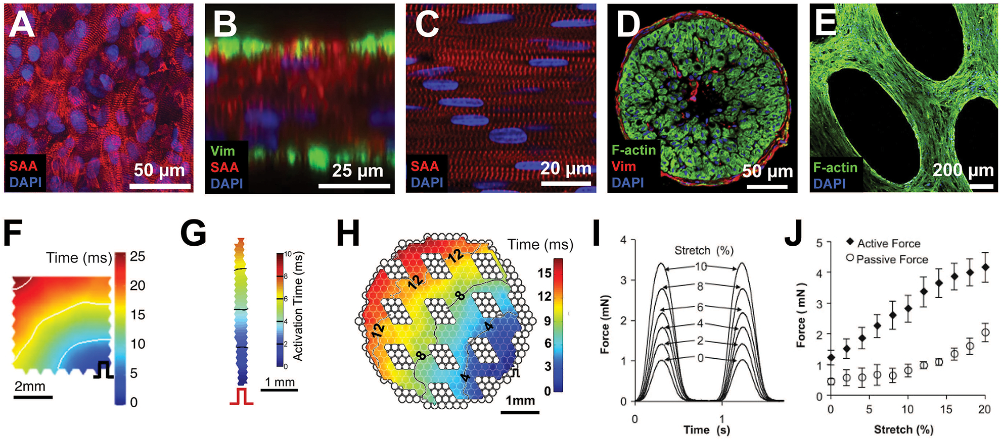
Figure 2 –

Functional properties of engineered cardiac tissues. Representative immunohistochemistry images of (A) a 7 × 7 mm hiPSC-CM patch, and (B) its cross section, (C) a 7 × 2 mm NRVM bundle and (D) its cross section, and (E) a 7 × 7 mm hESC-CM network patch. Representative isochrone activation maps from (F) a 7 × 7 mm hiPSC-CM patch, (G) a 7 × 2 mm NRVM bundle, and (H) a 7 × 7 mm mESC-CM network patch. (I) Representative force traces during stretch of a 1 Hz electrically stimulated 7×7 mm hESC-CM cardiac network patch. (J) Active and passive force-length relationships for 7×7 mm hESC-CM cardiac network patches. (Figure contains elements reproduced from references [18,2,21,20] with permission) (SAA = sarcomeric alpha actinin; Vim = vimentin)
