
FIGURE 1.
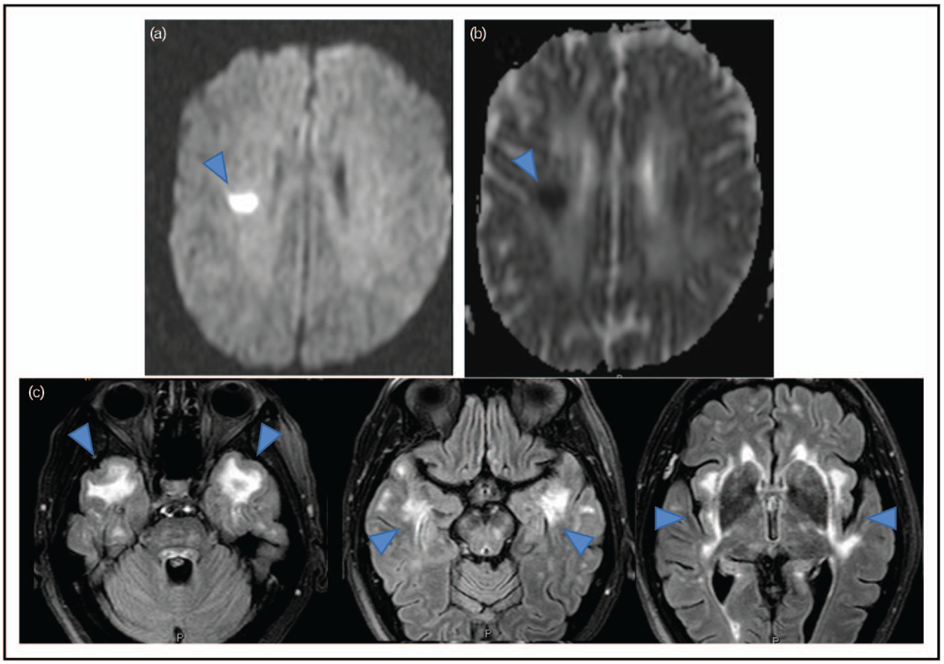
Typical Cerebral Autosomal Dominant Arteriopathy with Subcortical Infarcts and Leukoencephalopathy (CADASIL) MRI findings: panels a and b show typical acute ischemic changes in a CADASIL patient after stroke. Panel c consists of three cross-sectional MRI images to show typical white matter changes in a CADASIL patient. Blue arrows point to the typical changes.