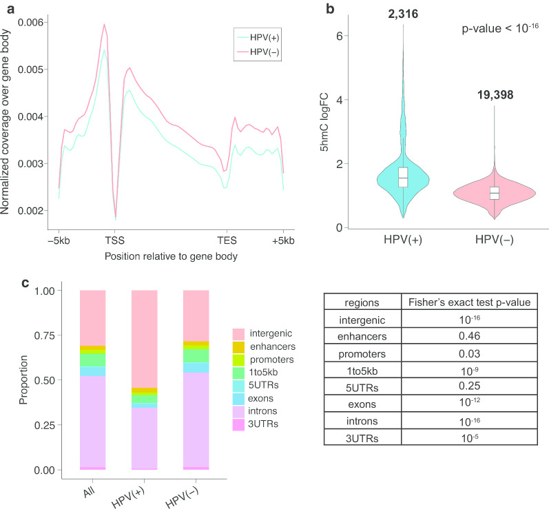
Fig. 1.
The level and distribution of hydroxymethylation varies by HPV status. a Global 5hmC distribution pattern over gene bodies in both HPV(+) and HPV(−) samples. b Violin plot of 5hmC logFC in HPV(+) tumors (left) and HPV(−) tumors (right). The number on top indicates the total number of peaks being tested. Wilcoxon signed-rank test p value < 10–16. c The distribution of hyper-5hmC peaks from HPV(+) and HPV(−) samples, where first column represents the combination of HPV(+) and HPV(−) tumors. The table on the right displayed the p values from Fisher’s exact test between HPV(+) and HPV(−) HNSCC, where exons and introns showed a p value of 10–12 and 10–16 respectively

