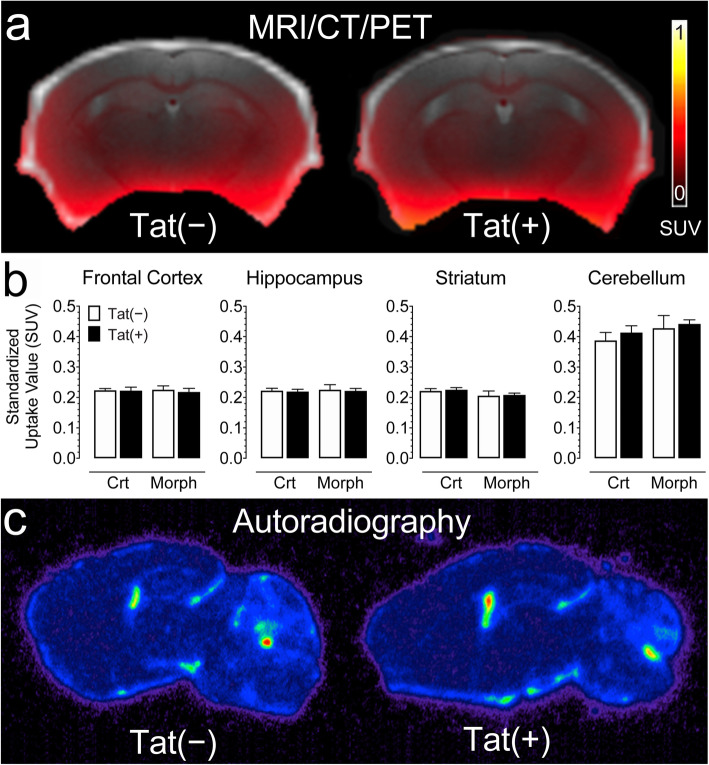
Fig. 3.
No significant changes were noted for neuroinflammation assessed by in vivo PET imaging using the [18F]-PRB111 probe in Tat transgenic mice in the presence or absence of morphine treatment. a A representative overlay image of a Tat(−) and Tat(+) mice show MR (gray), CT (white), and PET (SUV scale, yellow-red) imaging in the coronal plane. b PET imaging of Tat transgenic mice demonstrates the successful uptake of the [18F]-PBR111 radiotracer in different brain regions. No significant effect was noted for drug and/or genotype. c A representative brain image of a Tat(−) and Tat(+) mouse shows similar distribution of [18F]-PBR111 binding via post-mortem autoradiography in both groups in the parasagittal plane approximately 2 mm from the midline. All data are expressed as mean ± SEM. Statistical significance was assessed by ANOVA followed by Tukey’s post hoc test. SUV standardized uptake value, Crt no injections, Morph short-term (8-day) morphine injections. n = 5–7 mice per group

