Figure 2.
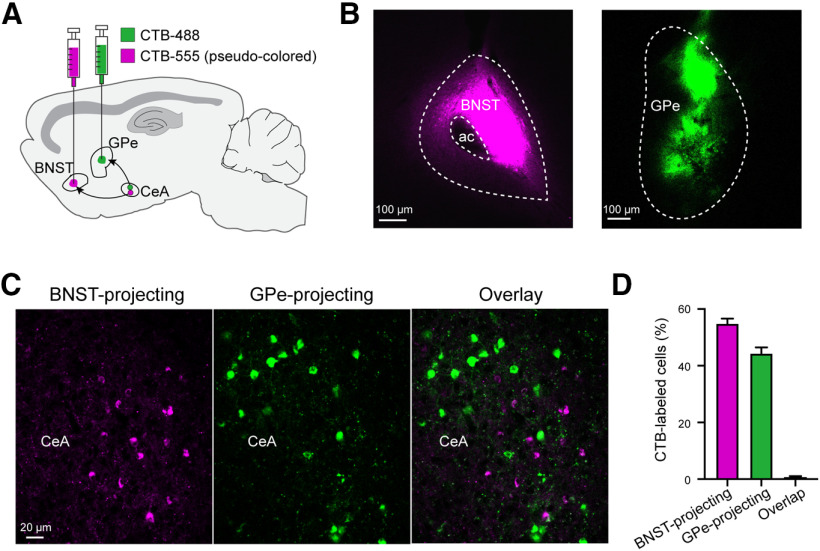
GPe-projecting CeA neurons do not send collateral projections to the BNST. A, Schematic of the approach. B, Histology images showing CTB-555 (pseudo-colored) and CTB-488 injection locations in the BNST and GPe, respectively, of a representative mouse. C, Representative confocal images of the CeA in the same mouse as that in B, showing CeA neurons labeled by CTB-555 and CTB-488. D, Quantification of the CeA neurons projecting to the BNST, GPe or both structures (n = 2 mice). Data in D is presented as mean ± SEM.

