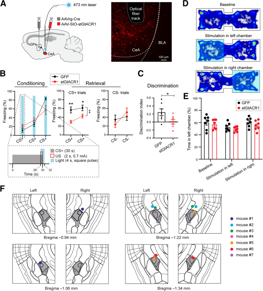
Figure 5.
GPe-projecting CeA neuron activity during US presentation is necessary for learning during fear conditioning. A, left, Schematic of the approach. Right, Representative confocal image showing the GPe-projecting CeA neurons expressing stGtACR1. The track of the implanted optic fiber is also shown. B, Freezing behavior in mice in which GPe-projecting CeA neurons expressed stGtACR1 (n = 7) or GFP (n = 7), during conditioning (left) and retrieval (right) sessions. Inset, Structure and timing of CS+, US, and light delivery. C, Discrimination Index calculated as [CS+ – CS–/[CS+ + CS–], where CS+ and CS– represent the average freezing during the presentation of CS+ and CS–, respectively. (D) Heat-maps for the activity of a representative mouse at baseline (top), or in a situation whereby entering the left (middle) or right (bottom) side of the chamber triggered photo-inactivation of GPe-projecting CeA neurons. E, Quantification of the mouse activity as shown in D, for mice in which stGtaCR1 (n = 7) or GFP (n = 7) was introduced into GPe-projecting CeA neurons. F, Schematics showing the placement of optic fibers in all the mice used for inhibiting GPe-projecting CeA neurons with optogenetics (n = 7 mice). The CeA is colored in dark gray. Data in B, C, E are presented as mean ± SEM. *p < 0.05, **p < 0.01.

