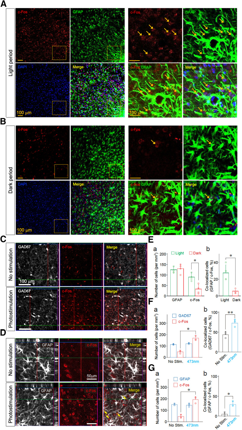
Figure 9.
The activity of VLPO astrocytes is increased during sleep states. A, B, c-Fos expression in the VLPO astrocytes during the light (sleep; A) and dark (wake; B) periods. Confocal images of c-Fos (red) and GFAP immunoreactivity (green) in the VLPO region. Frozen sections of brain were prepared from animals in sleep (light period: 2–4 P.M.) and wake (dark period: 7–9 P.M.) states. Colocalizations of c-Fos (red) and GFAP immunoreactivity (green) in the VLPO region are indicated by arrows. Cell nuclei were stained with DAPI to confirm the nuclear expression of c-Fos. C, D, Increased c-Fos immunoreactivity in GABAergic neurons and astrocytes within the VLPO region following optogenetic stimulation. Rats were injected with AAV-ChR2-eYFP (n = 3). Frozen sections of brains were prepared 60 min after the animals received photostimulation. Neuronal and astrocytic expression of c-Fos in the VLPO area was identified using anti-GAD67 (C) and anti-GFAP (D) antibodies, respectively, in conjunction with anti-c-Fos antibodies. Cell nuclei were stained with DAPI to confirm the nuclear expression of c-Fos. GAD67-double positive and c-Fos-double positive cells or GFAP-double positive and c-Fos-double positive cells are indicated by arrows. E, Quantification of immunopositive cells for GFAP and c-Fos (a) and their co-localization (b). Frozen sections of brain were prepared from animals in sleep (light period: 2–4 P.M.) and wake (dark period: 7–9 P.M.) states. Astrocytic expression of c-Fos in the VLPO region was identified using an anti-GFAP antibody. Each column and error bar represents the mean and SD from four experiments; *p < 0.05; unpaired t test; c-Fos positive cells, t(6) = 2.53, p = 0.0223; co-localized cells, t(6) = 2.98, p = 0.0122. F, Quantification of GAD67 and c-Fos immunoreactive cells (a) and their co-localization (b). Rats were injected with AAV-ChR2-eYFP (n = 3). Frozen sections of brain were prepared 60 min after animals received photostimulation. Neuronal expression of c-Fos in the VLPO region was identified using an anti-GAD67 antibody. Each column and error bar represents the mean and SD from three experiments; *p < 0.05; **p < 0.01; unpaired t test; c-Fos-positive cells, t(4) = 2.84, p = 0.0233; co-localized cells, t(4) = 3.765, p = 0.0098. G, Quantification of immunopositive cells for GFAP and c-Fos (a) and their co-localization (b). Rats were injected with AAV-ChR2-eYFP (n = 3). Frozen sections of brain were prepared 60 min after the animals received photostimulation. Astrocytic expression of c-Fos in the VLPO region was identified using an anti-GFAP antibody. Each column and error bar represents the mean and SD from three experiments; *p < 0.05; unpaired t test; c-Fos-positive cells, t(4) = 2.96, p = 0.0206; co-localized cells, t(4) = 3.54, p = 0.0119.

