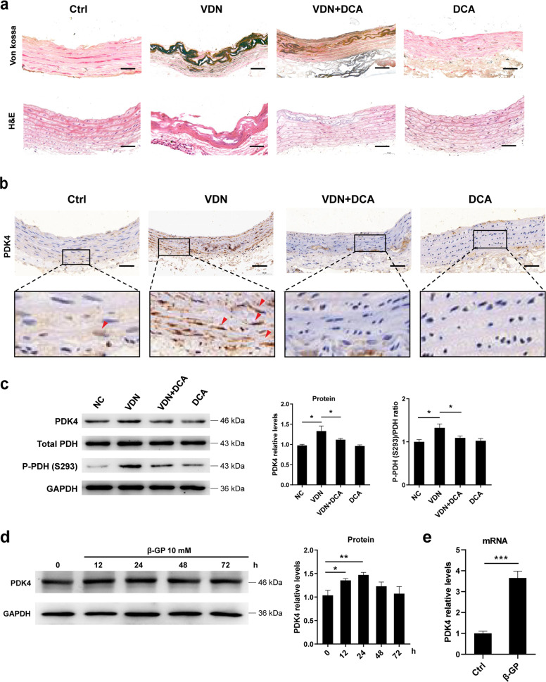
Fig. 1. PDK4 is upregulated during the vascular calcification.
a Representative Von Kossa staining and H&E staining in aortic tissues isolated from SD rats (original magnification, ×200; Scale bars = 100 μm). N = 6 rats per group. b Representative immunohistochemical staining of PDK4 in aortic tissues isolated from SD rats (original magnification, ×200; Scale bars = 100 μm). N = 6 rats per group. c Protein expression of PDK4, phospho-PDH (S293), and PDH was determined by western blotting. N = 4–6 rats per group. d VSMCs were exposed to 10 mM β-GP for 0–72 h, and the protein expression of PDK4 was determined using western blotting. N = 3 independent experiments. e VSMCs were treated with 10 mM β-GP for 24 h. RT-qPCR analysis was used to detect the mRNA level of PDK4 in the VSMCs. N = 3 independent experiments. *P < 0.05, **P < 0.01, ***P < 0.001.

