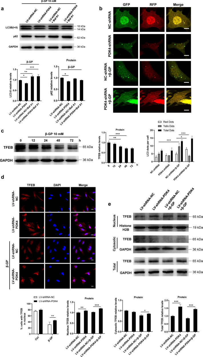
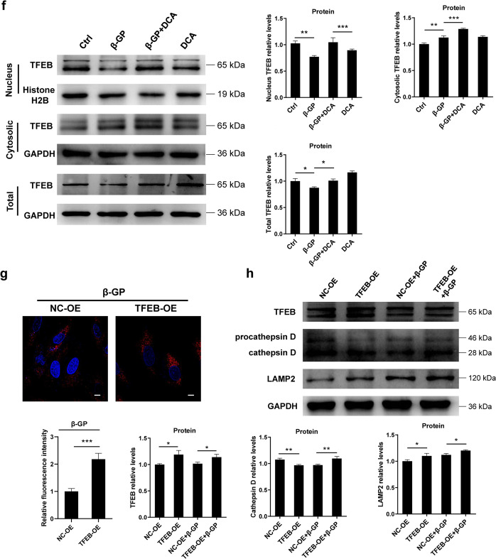
Fig. 4. Inhibition of PDK4 promotes autophagy activity.
a VSMCs were transfected with lentivirus carrying PDK4 shRNA and then exposed to β-GP for 24 h. The protein expression levels of LC3 and p62 were measured by western blotting. N = 3 independent experiments. b Confocal microscopy of the lentivirus-mRFP-GFP-LC3-transduced VSMCs. Scale bars: 10 μm, N = 40–50 cells per group. c VSMCs were treated with 10 mM β-GP for 24 h, and TFEB protein expression was determined by western blotting. N = 3 independent experiments. d VSMCs were transfected with shRNA targeting PDK4 for 48 h. Then, the cells were incubated in the presence or absence of β-GP. After 24 h, the VSMCs were stained with an antibody against TFEB and analysed by confocal microscopy. Scale bars: 10 μm, N = 40–50 cells per group. e Western blotting analysis of the total, nuclear, and cytoplasmic fractions from PDK4-shRNA-transfected VSMCs in the presence or absence of β-GP. N = 3 independent experiments. f Cells were treated with 1 mM DCA in the presence or absence of β-GP for 24 h. TFEB protein levels in the total, cytosolic and nuclear fractions were determined by western blotting. N = 3 independent experiments. g The lysosome content of the VSMCs was probed with LysoTracker Red, and images were taken under a confocal microscope. Scale bars = 10 μm. N = 40–50 cells per group. h Western blotting analysis of LAMP2, TFEB and Cathepsin D expression in the calcified VSMCs transfected with a plasmid containing the TFEB gene. N = 3 independent experiments. *P < 0.05, **P < 0.01, ***P < 0.001.


