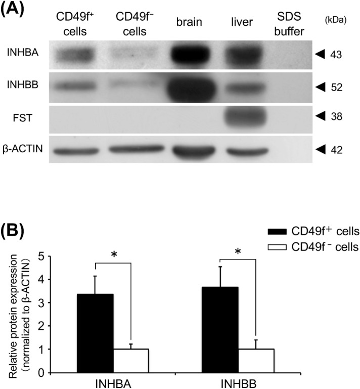
Figure 2.

(A) Western blot analysis for freshly-isolated cells: Proteins of INHBA, INHBB, and follistatin (FST) were detected from protein extracts (30 μg/lane). β-actin was used as an internal control by re-probing. Brain protein extracts, positive control of INHBA and INHBB; liver protein extracts, positive control of FST; SDS buffer, negative control. Typical blotting images are shown from 3 independent experiments. One set of experiments was performed using protein extracts of CD49f+ and CD49f- cells fractionated from the salivary glands of 3 mice, and 3 sets of experiments carried out independently. (B) Densitometric analysis for protein levels of INHBA, INHBB, and FST. Signals were normalized to β-actin. One set of experiments was performed using protein extracts of CD49f+ and CD49f- cells fractionated from the salivary glands of 3 mice, and 3 sets of experiments carried out independently. *P < 0.05, Student’s t test.
