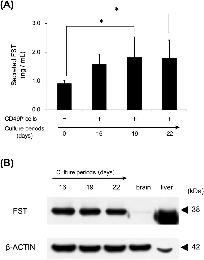
Figure 5.
CD49f+ cells were cultured for 16, 19, and 22 days, and the supernatants and intracellular proteins were used in the following assays. (A) FST levels in the supernatants of cultured CD49f+ cells. FST amount at a specific culture period was measured by using ELISA. The experiment was performed using protein extracts of sub-cultured CD49f+ cells fractionated from the salivary glands of 3 mice, and 3 independent experiments were carried out. *P < 0.05, one-way ANOVA and Tukey’s HSD test. (B) Intracellular FST level in cultured CD49f+ cells. FST amount at specific culture period was detected by using western blotting (30 μg/lane). β-actin was used as an internal control by re-probing. Brain protein extracts and liver protein extracts were used as positive controls. Typical blotting images are shown in 3 independent experiments, and the experiment was performed using sub-cultured CD49f+ cells fractionated from the salivary glands of 3 mice.

