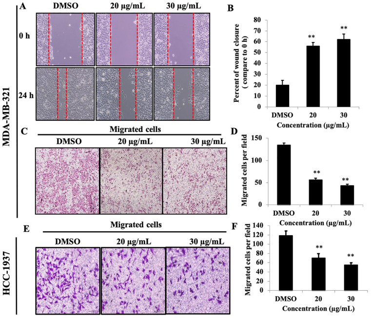
Figure 2.
PEF-III attenuates wound closure and cell migration of TNBC cells. Cell monolayers were scraped with a sterile p200 pipette tip, and the cells were treated with the indicated concentrations of PEF-III for 24 hours. The wound closure distances were quantified by measuring the distance between the edges (A and B). PEF-III also inhibited cell motility (C and E), as determined by a transwell cell migration assay. The migrated cells were stained with crystal violet, and photographs were taken for the inhibition calculation (D and F).*
The results are presented as the means ± SDs of 3 independent experiments. Bars with asterisks indicate significant differences from the control at P ≤ .05 (*) or P ≤ .01 (**).

