Fig. 3.
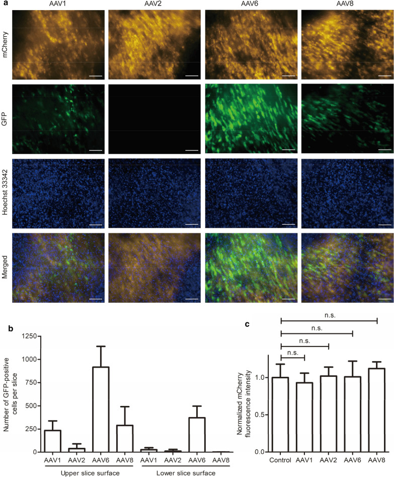
Gene transfer efficiency, penetration depth, and cytotoxicity of AAV1, 2, 6, and 8. a Myocardial slices were infected with four different AAV serotypes. After 5 days, slices were stained with Hoechst 33342 to label nuclei. AAV-mediated GFP expression was analyzed by fluorescence microscopy; mCherry-positive cells indicate CMs. The images shown represent the upper slice side. Scale bars, 100 µm. b Enumeration of GFP-positive cells per slice. Images were taken from both the upper and the lower slice side on day 5 after infection and GFP-positive cells were counted. c AAV cytotoxicity. mCherry signal was measured on day 5 after AAV infection on both slice surfaces and normalized to the mCherry fluorescence intensity of slices without virus (control). In b and c, signals of three independent experiments with nine slices in total were quantified, and means and SDs (error bars) are given. n.s. indicates not significant

