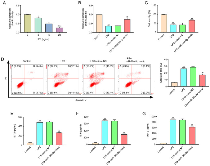
Figure 4.
Overexpression of miR-26a-5p decreases apoptosis and inflammatory responses in LPS-induced A549 cells. A549 cells were transfected with miR-26a-5p mimic or mimic NC (10 nM), and then treated with LPS for 24 h, followed by the assessment of cell apoptosis and inflammatory response. (A) The expression of miR-26a-5p was detected by reverse transcription-quantitative PCR in A549 cells treated with various concentrations (0, 5, 10 and 20 µg/ml) of LPS. (B) The expression of miR-26a-5p was detected in transfected A549 cells. (C) The cell viability was measured by MTT assay in transfected A549 cells. (D) Apoptotic cells were detected by flow cytometry in transfected A549. The expression levels of (E) IL-1β, (F) IL-6 and (G) TNF-α were detected by ELISA in transfected A549 cells. Data were presented as the mean ± SD. (A) *P<0.05, **P<0.01 vs. LPS (0 µg/ml) group. (B-G) **P<0.01 vs. Control group; ##P<0.01 vs. LPS + mimic NC group. miR, microRNA; NC, negative control; LPS, lipopolysaccharide.

