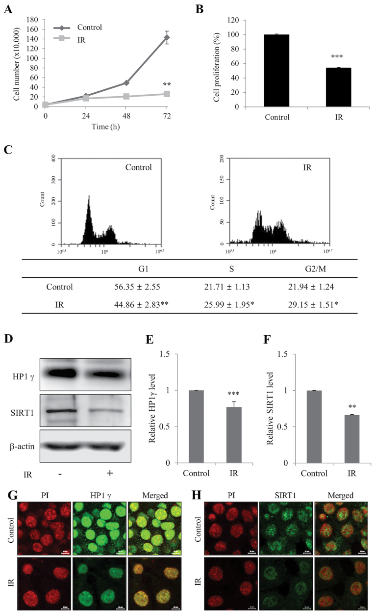
Figure 1.
Radiation induces senescence of mIMCD-3 cells. (A) Control and IR-treated viable mIMCD-3 cells were visualized using Trypan blue exclusion and counted. (B) Control and IR-treated cell proliferation wase determined using the WST assay. Data are presented as a percentage of control cell growth. (C) Control and IR-treated mIMCD-3 cells were stained with PI and subjected to flow cytometry. (D) Protein expression levels of HP1γ and SIRT1 were determined via western blotting, with β-actin as the loading control. Semi-quantification of (E) HP1γ and (F) SIRT1 protein expression levels in cell lysates relative to the control group. Immunofluorescence analysis of (G) HP1γ and (H) SIRT1 using a fluorescein isothiocyanate-conjugated secondary antibody and PI nuclear staining followed by confocal microscopy (scale bar, 10 µm). mIMCD-3 cells were treated with or without 6 Gy radiation and cultured for 72 h. Data are expressed as the mean ± SD (n=4), *P<0.05, **P<0.01, ***P<0.001 vs. Control. mIMCD-3, mouse inner medullary collecting duct-3; IR, ionizing radiation; HP1γ, heterochromatin protein 1γ; SIRT1, sirtuin 1; PI, propidium iodide.

