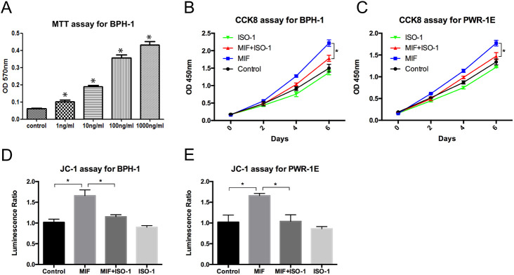
Fig. 2.
MIF promoted proliferation of BPH-1 and PWR-1E cells. (A) MTT assay showed the proliferation of BPH-1 cells treated with various concentrations of rMIF for 3 days. Data are presented as mean±s.d., n=3. (B) CCK8 assay showed the proliferation of BPH-1 cells treated with control, rMIF (100 ng/ml), rMIF (100 ng/ml)+ISO-1 (10 µM) and ISO-1 (10 µM), respectively. Data are presented as mean±s.d., n=3. (C) CCK8 assay showed the proliferation of PWR-1E cells treated with control, rMIF, rMIF+ISO-1 and ISO-1, respectively. Data are presented as mean±s.d., n=3. (D) JC-1 assay showed the growth rates of the BPH-1 cells treated by control, rMIF, rMIF+ISO-1 and ISO-1, respectively. Data are presented as mean±s.d., n=3. (E) JC-1 assay showed the growth rates of the PWR-1E cells treated by control, rMIF, rMIF+ISO-1 and ISO-1, respectively. Data are presented as mean±s.d., n=3. *P<0.05, *P<0.05. Statistical analyses were performed using ANOVA followed by Tukey's test for multiple comparison.

