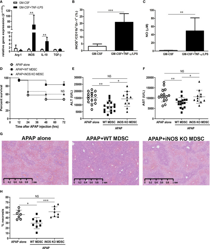
Figure 6.
TNF-α/lipopolysaccharide (LPS) myeloid-derived suppressor cells (MDSCs) ameliorate acetaminophen (APAP)-induced liver injury through iNOS (A) mRNA expression of in vitro-generated MDSCs cultured with GM-CSF alone or combined with TNF-α/LPS. (n=6),**P < 0.01, Mann-Whitney test. (B) iNOS expression in MDSCs was measured by flow cytometry(n=8), ***P < 0.001, Mann-Whitney test. (C) NO production in the culture media of MDSCs (n=6), **P < 0.01, Mann-Whitney test. (D) BALB/c mice were adoptively transferred with WT or iNOS KO TNF-α/LPS MDSCs just after APAP challenge. The survival of mice was plotted (n=17-20). *P < 0.05, NS, no significance, Log-rank test. (E) The serum ALT at 12 h after APAP challenge (n=11-16). (F) The serum AST at 12 h after APAP challenge (n=11-16).*P < 0.05, **P < 0.01, NS, no significance. Kruskal-Wallis test. Data were expressed as mean ± SD. (G) Representitive of liver histological stain of mice with indicative treatment. (H) The quatification of liver necrosis (n=7-8). *P < 0.05, ***P < 0.001, NS, no significance. Kruskal-Wallis test. Data were expressed as mean ± SD.

