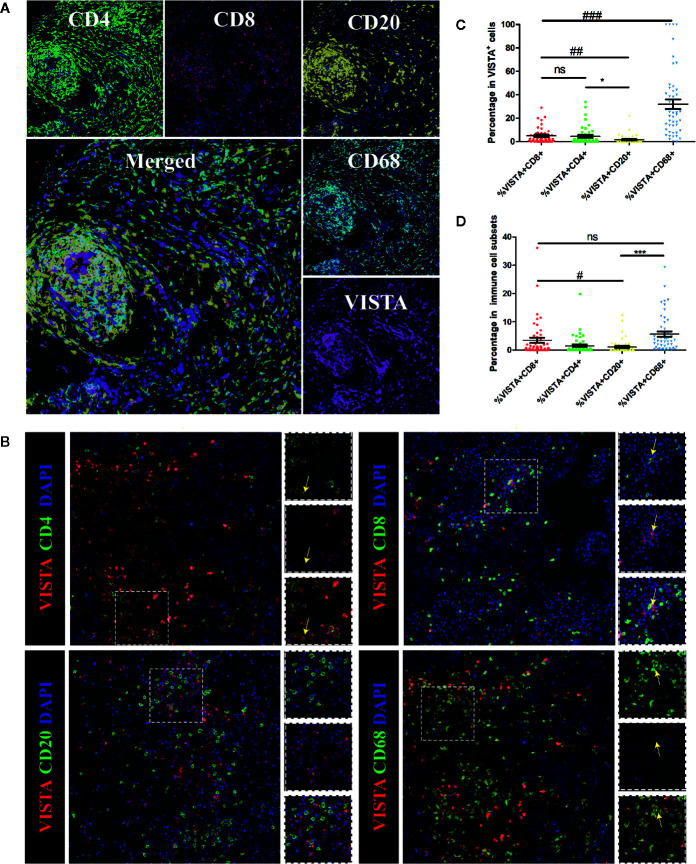
Figure 4.
Multiplex immunofluorescence for VISTA and selected tumor-infiltrating immune cell markers in human breast cancer tissue. Markers of tumor-infiltrating immune cells: CD68 (macrophages), CD4 (T cells), CD8 (cytotoxic T cells) and CD20 (B cells). (A) Representative images of biomarkers in human breast cancer tissue. CD4 staining is shown in green; CD8 staining is shown in red; CD20 staining is shown in yellow; CD68 staining is shown in cyan; VISTA staining is shown in magenta; and DAPI staining is shown in blue. 400×. (B) Co-localization of VISTA with selected tumor-infiltrating immune cell markers in breast cancer detected by immunofluorescence. CD4, CD8, CD20, and CD68 staining is shown in green; VISTA staining is shown in red; and DAPI staining is shown in blue. Areas of co-localization are indicated with yellow arrows. 400×. (C). Summary plot of the proportion of each subpopulation of cells among double-positive cells. Each dot represents data from an individual patient. P-values were obtained by an unpaired T-test. * p < 0.05; ## p < 0.01; ### p < 0.001. (D) Summary plot of the proportion of double-positive cells in each subpopulation of cells. Each dot represents data from an individual patient. P-values were obtained by an unpaired T-test. # p < 0.05; *** p < 0.001. NS, no significance.

