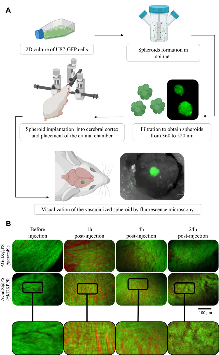
Figure 3.
Vascular selectivity using the cranial chamber model with U87-GFP spheroid and fluorescence imaging. (A) Experimental protocol illustrating spheroids formation (U87-GFP cells grown in non-adherent flasks were transferred to a spinner placed on a magnetic stirrer for 4 days to form spheroid), their filtration and implantation onto the cerebral cortex of nude mice. A glass window was placed on the skull allowing visualization of the tumor tissue and its vasculature at about 10 days after xenotransplant. Mice were divided into two batches: AGuIX@PS@KDKPPR or AGuIX@PS@scramble group. Visualization of different nanoparticles into the vasculature was performed on anesthetized mice. Blood vessels are observed in black and PS fluorescence in red. (B) The vascular network was imaged before, 1, 6 and 24h after i.v. injection of nanoparticles (4 μmol.kg−1, porphyrin equivalent). The excitation filter was at 560 ± 25 nm and at 475 ± 20 nm for PS and GFP-U87 fluorescent images acquisition, respectively. The emission filter was composed with a block of four emission filters at 440, 521, 607 and 700 nm. Photos taken with the same magnification; scale bar represents 100 µm.
Abbreviations: GFP, green fluorescent protein; i.v., intravenous; PS; photosensitizer.

