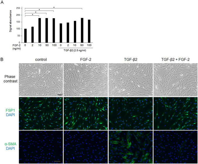
Fig 3. Effects of FGF-2 on cell proliferation (A), cell morphology, and expressions of cell-type markers (B).
Conjunctival fibroblasts were treated with FGF-2 (0–100 ng/mL) in the presence or absence of 2.5 ng/mL TGF-β2 for 48 h. Subsequently, cells were observed by phase contrast microscopy, and were subjected to the WST-8 assay or immunofluorescence assay. Both FGF-2 and TGF-β2 increased cell proliferation without an additive effect (A). TGF-β2 treatment induced some changes in cell shape (from spindle- to cobblestone-like); FGF-2 did not affect cell shape in the presence or absence of TGF-β2 (B). At all conditions, cells were FSP-1 positive, while α-SMA expressed only in the cells treated with TGF-β alone. *p < 0.05. Data are shown as the mean ± SE, n = 8 for WST-8 assay, n = 3 for immunofluorescence assay. Scale bar: 200 μm.

