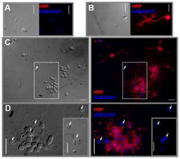
Figure 1. Dissociated CNS cells displayed neuritic outgrowth and were horseradish peroxidase-(HRP)-positive.

Dissociated central nervous system (CNS) cultures (2CNS/corverslip) were incubated for 1 day, immunostained with anti-HRP, and the nuclei stained with Hoechst. Left panels show the corresponding light picture. (A) Negative control for anti-HRP (exposed only to the secondary antibody: Alexa-Fluor-546-Goat-Anti-Rabbit). (B-D) Positive labeling with anti-HRP. The main panel in (D) corresponds to the inset area in (C) that was rotated by 90°. The inset in (D) corresponds to another field that shows a group of HRP-negative cells. The arrows indicate some of the HRP-negative cells. (63x objective, bar=10μm).
