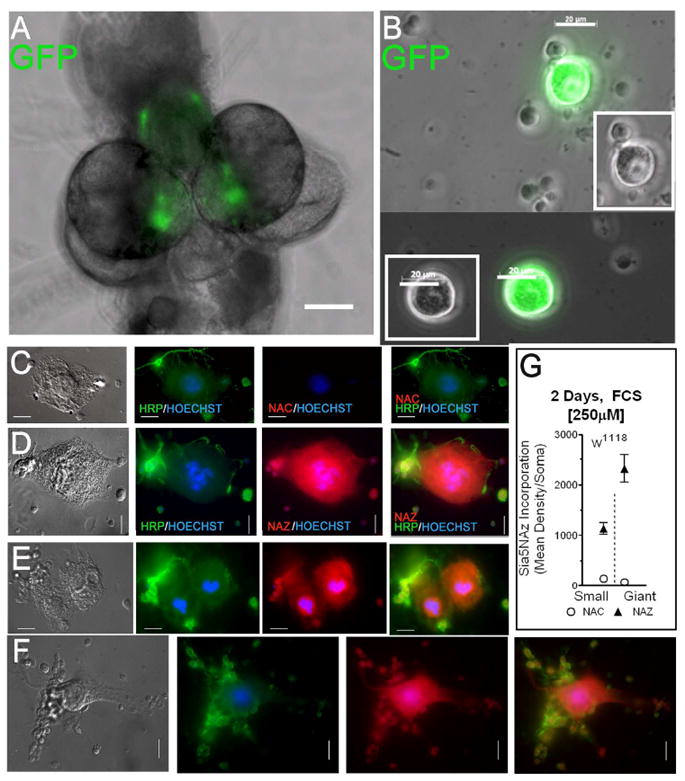
Figure 4. Cultured giant neurons incorporate sialic acids.

(A,B) Identification of giant neurons in third instar larvae in whole CNS (10x objective, bar=100μm) (A) and dissociated CNS (40x objective, bar=20μm) (B). (A,B) The giant neurons were labeled with membrane-targeted green fluorescent protein (GFP) by using UAS-mc8-GFP driven by OK307-Gal4. (C-F) Show pictures of giant and smaller CNS neurons derived from w1118 Dm. Cells (5 CNS/coverslip) were cultured for 2 days in medium containing heat-inactivated serum and 250μM of either NAC (C) or NAZ (D-F). Cells were immunostained with anti-HRP; and stained with the Alexa-Fluor-546-Alkyne and Hoechst for nuclei. (63x objective, bar=10μm). (G) Shows the quantification of Sia5NAz incorporation in giant neurons (n=7) as compared to that of close by smaller neurons (n=34) (mean ± sem).
