
Fig. 6. DEL-1 deficiency promotes neutrophil recruitment to perimetastatic foci in the lung.
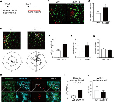
(A) Scheme of two-photon intravital imaging. DsRed-B16F10 cells were intravenously injected (day 0) and imaged at day 5 for GFP+ neutrophils and DsRed+ B16F10 cells. (B and C) Representative images (B) and quantification (C) of neutrophils around metastatic foci in the lungs of LysM-GFP/WT (n = 10) or LysM-GFP/Del1 KO mice (n = 7). Scale bar, 50 μm. (D) Migration trajectories of neutrophils observed for 15 min in a field of view. The starting point for each neutrophil was set to zero. Axis unit = μm. (E to G) Quantification of neutrophil velocity (E), displacement (F), and meandering index (G) from intravital imaging. (H) Broad view of cleared lung tissue 5 days after DsRed-B16F10 cell injection. Scale bar, 300 μm. (I) Counts of neutrophils within 50 μm from the boundaries of metastatic foci. (J) The number of neutrophils inside the metastatic foci. *P < 0.05; ****P < 0.0001.