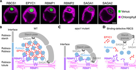
Fig. 4. The motif orchestrates the architecture of the pyrenoid’s three subcompartments.

(A) Representative confocal images of Venus-tagged proteins that have the Rubisco-binding motif and Rubisco small subunit (RBCS1). Chlorophyll autofluorescence delimits the chloroplast. Scale bar, 2 μm. (B) Proposed model for how the motif mediates assembly of the pyrenoid’s three subcompartments in wild type. The motif on tubule-localized transmembrane proteins RBMP1 and RBMP2 mediates Rubisco binding to the tubules [Retic. region is the reticulated region of the tubules (9)]. Multiple copies of the motif on EPYC1 link Rubiscos to form the pyrenoid matrix (21). At the periphery of the matrix, the motif on starch-binding proteins SAGA1 and SAGA2 mediates interactions between the matrix and surrounding starch sheath. (C) The model in (B) explains the matrix-less phenotype observed in EPYC1-less mutants. (D) The model also explains the absence of matrix and starch plates in mutants where Rubisco’s binding site for the motif has been disrupted (21).
