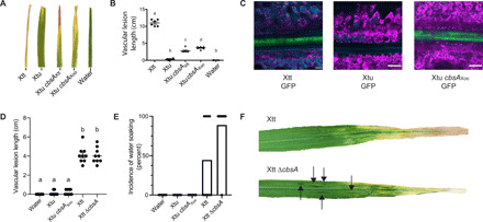
Fig. 2. Experimental gain and loss of CbsA facilitates transitions between vascular and nonvascular pathogenic lifestyles.

(A) Addition of either cbsA from vascular X. translucens pv. translucens (Xtt) or cbsA from vascular X. oryzae pv. oryzae (Xoo) to nonvascular X. translucens pv. undulosa (Xtu) permits development of chlorotic lesions indicative of vascular disease on barley 21 days post-inoculation (dpi). (B) Corresponding vascular lesion lengths, with significant differences among treatments indicated by a to d (n = 6, P < 0.02). (C) Representative confocal images of vascular bundles downstream of leaf lesions on barley 12 dpi with GFP transformed strains demonstrate gain of vascular colonization by Xtu cbsAXoo. Green indicates bacterial cells expressing GFP; magenta indicates chlorophyll autofluorescence outlining nonvascular mesophyll cells; cyan indicates autofluorescence outlining xylem cell walls or phenylpropanoid accumulation in mesophyll cells. (D and E) Lesion lengths or incidence of nonvascular water-soaked lesions were quantified after barley leaf clipping 14 dpi with Xtt ∆cbsA. Bars in (E) represent percent leaves showing symptoms with dots included to display individual leaf lesion incidence. (F) Images of symptomatic barley leaves infected with Xtt and Xtt ∆cbsA, where water-soaked lesions are indicated, with black arrows indicating nonvascular symptom development.
