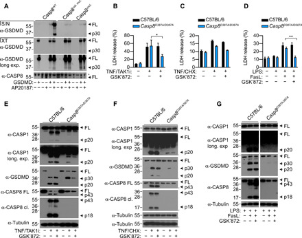
Fig. 4. Caspase-8 dimerization and autoprocessing is required for GSDMD cleavage.

(A) HEK 293T cells were transfected with GSDMD together with WT caspase-8 (Casp8WT), caspase-8 catalytic mutant (Casp8cat. mut.), or uncleavable caspase-8 (Casp8uncl.). Vector control was transfected such that each set of constructs received equivalent amounts of DNA. Twenty-four hours after transfection, transfected cells were treated with AP20187 (dimerizer) for a further 6 hours. Cell extracts (XT) and precipitated supernatants (S/N) were analyzed by immunoblotting for GSDMD and caspase-8. (B to G) WT or caspase-8 uncleavable (D387A) Casp8D387A/D387A BMDMs were stimulated with TNF (100 ng/ml)/TAK1i (125 nM) or TNF (100 ng/ml)/CHX (10 μg/ml) or primed with LPS (100 ng/ml) for 16 hours and treated with FasL (100 ng/ml). Where indicated, cells were treated with GSK’872 (1 μM) 20 to 30 min before stimulation. LDH release was measured at (B and C) 4 hours or (D) 6 hours after stimulation. (E to G) Mixed supernatant (S/N) and cell extracts were examined by immunoblotting. (G) Blots are cropped from the same film. (E to G) Immunoblots are representative of three independent experiments. (B to D) Data are means + SEM of pooled data from four independent experiments. Data were normally distributed and analyzed using a parametric t test. *P < 0.05 or **P < 0.01.
