
Fig. 2. Disque Platform.
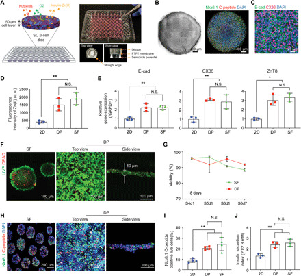
(A) Scheme and gross view of 3D reconstructed SC β cells in Disque Platform (DP). (B) Cell disc morphology shown through bright-field and confocal microscopy [NK6 homeobox 1 (Nkx6.1) and C-peptide]. (C) Immunofluorescent staining of E-cadherin (E-cad) and connexin 36 (CX36) in cell discs. (D) 3D cultures (DP and SF) showed significantly higher levels of Zn(II) compared to conventional 2D system (2D monolayer), measured through signal intensity from FluoZin-3 indicator dye (n = 3, **P < 0.01 versus 2D). N.S., not significant; a.u., arbitrary units. (E) SC β cells cultured in DP show significantly higher gene expression of E-cad, CX36, and zinc transporter 8 (ZnT8) using quantitative real-time polymerase chain reaction analysis (n = 3, *P < 0.05, **P < 0.01 versus 2D). (F) Homogenous distribution of viable pancreatic progenitor (PP) cells in DP via LIVE/DEAD assay (5-day differentiation). (G) DP achieved high percentage of viable cells comparable to SF culture during PP differentiation to SC β cells [stage 4 day 1 (S4d1) to S6d7)] via flow cytometry analysis with the Zombie Aqua Fixable Viability Kit (n = 3). (H) DP shows similar expression of C-peptide and Nkx6.1 in SC β cells to SF through immunocytochemistry staining and (I) flow cytometry analysis (n = 4, **P < 0.01 versus 2D). (J) SC β cells show similar glucose stimulated insulin secretion index between DP and SF (n = 3, **P < 0.01 versus 2D).