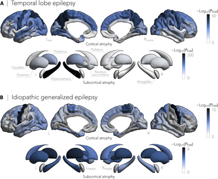
Fig. 1. Cortical thickness and subcortical volume in TLE and IGE.

(A) Cortical thickness and subcortical volume reductions in TLE (n = 732), compared to healthy controls (n = 1418), spanned bilateral precuneus (PFDR < 4 × 10−36), precentral (PFDR < 8 × 10−36), paracentral (PFDR < 6 × 10⁻²⁹), and superior temporal (PFDR < 3 × 10⁻¹⁴) cortices and ipsilateral hippocampus (PFDR < 2 × 10−199) and thalamus (PFDR < 5 × 10−64). (B) In contrast, gray matter cortical and subcortical atrophy in IGE (n = 289), relative to controls (n = 1075), was more subtle and affected predominantly bilateral precentral cortical regions (PFDR < 9 × 10⁻¹⁰) and the thalamus (PFDR < 3 × 10⁻⁶). Negative log10-transformed FDR-corrected P values are shown.
