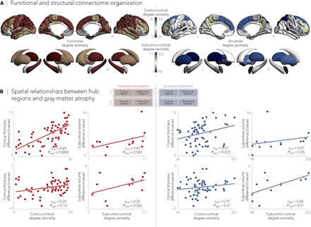
Fig. 2. Epilepsy-related atrophy correlates with hub organization.

(A) Normative functional and structural network organization, derived from the HCP dataset, was used to identify hubs (i.e., regions with greater degree centrality). (B) Schematic of the figure layout is pictured in the middle. Gray matter atrophy related to node-level functional (left) and structural (right) maps of degree centrality, with greater atrophy in hub compared to nonhub regions. Stratifying findings across TLE and IGE, we observed stronger associations between cortico-cortical functional hubs and cortical atrophy patterns in TLE (Pspin < 0.0001) and between subcortical volume loss and subcortico-cortical structural hubs in IGE (Pshuf < 0.01).
