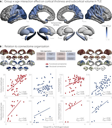
Fig. 4. Negative effects of age on cortical thickness and subcortical volume in TLE.

(A) Significant age-related differences on gray matter atrophy between individuals with TLE and healthy controls for all cortical and subcortical regions. Patients with TLE showed a negative effect of age on cortical thickness in bilateral temporo-parietal (PFDR < 0.005) and sensorimotor (PFDR < 0.01) cortices and on subcortical volume in ipsilateral hippocampus (PFDR < 5 × 10⁻⁷) and bilateral thalamus (PFDR < 0.05). Negative log10-transformed FDR-corrected P values are shown. (B) Schematic of the figure layout is provided in the middle. Scatterplots depict relationships between the age-related effects and functional (red) and structural (blue) maps of degree centrality (left) and disease epicenter (right). Significant associations were observed between age-related effects and every hub and epicenter measures, with the exception of structural subcortical degree centrality, suggesting a role of connectome organization on age-related effects in TLE.
