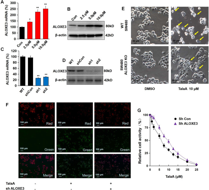
Fig. 6. TalaA enhanced ferroptosis in CRC cells by up-regulation of ALOXE3.
A ALOXE3 mRNA was increased by TalaA dose-dependently; *p < 0.05, **p < 0.01, N = 3 independent repeats. B The protein level of ALOXE3 was elevated by TalaA in a dose-dependent manner. C The mRNA level was decreased via lenti-shALOXE3 infection. **p < 0.01 versus ShCon, N = 3 independent repeats. D The ALOXE3 protein level was reduced by lenti-shALOXE3. E Although 10 μM TalaA violently caused cell membrane destroy in wild type SW480 cells, same concentration TalaA only led to mild membrane destroy in ALOXE3 knocked down SW480 cells. The yellow arrows indicated broken cells. F The lipid peroxidation was detected by cell-based lipid peroxidation assay kit. The stained cells were recorded with a fluorescence microscope. When the lipids were peroxidized, the fluorescence shifted from red to green. G The cell activity curve right shifted as ALOXE3 was knocked down. The black points represented wild type SW480, and purple triangles represented ALOXE3 knocked down SW480 cells. For each concentration point, three repeats were performed.

