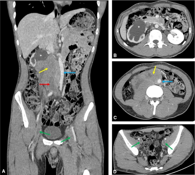
Figure 1.
Axial (B–D) and coronal (A) delayed phase (120 s) CT scan of the abdomen and pelvis demonstrating the right-sided retroperitoneal mass (yellow arrows) encasing the IVC (red arrow). The aorta is displaced to the right (blue arrow) and extensive thrombus is seen in the iliac veins (green arrows) and lower IVC. Severe right-sided hydronephrosis (asterisks) with associated cortical thinning also noted. IVC, inferior vena cava.

