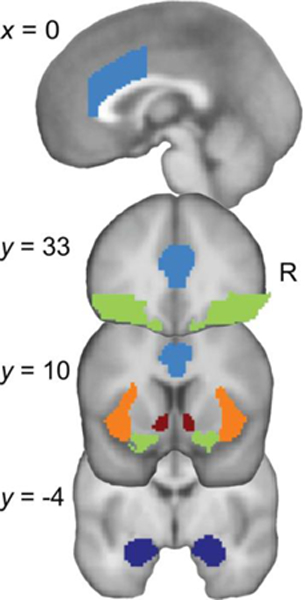
Fig. 2. Anatomical regions of interest (ROIs).

ROIs in the left and right dorsal anterior cingulate cortex (dACC, light blue), ventrolateral prefrontal cortex (vlPFC, green), anterior insula (AI, orange), nucleus accumbens (NAcc, red), and amygdala (AMY, dark blue), superimposed on T1-weighted template brain image (Montreal Neurological Institute). Top image shows a sagittal section, bottom 3 images show coronal sections. R, right
