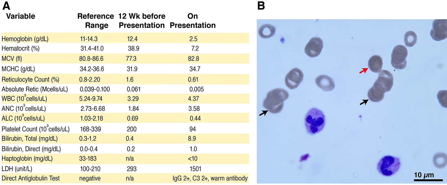
Figure 1: Laboratory evidence of autoimmune hemolytic anemia.

A) Panel A shows hematological parameters prior to presentation and upon admission. B) Panel B displays a representative image of the peripheral blood smear, demonstrating rouleaux formation (black arrows), presence of microspherocytes (red arrow), hypochromic microcytic red blood cells, as well as large platelets.
