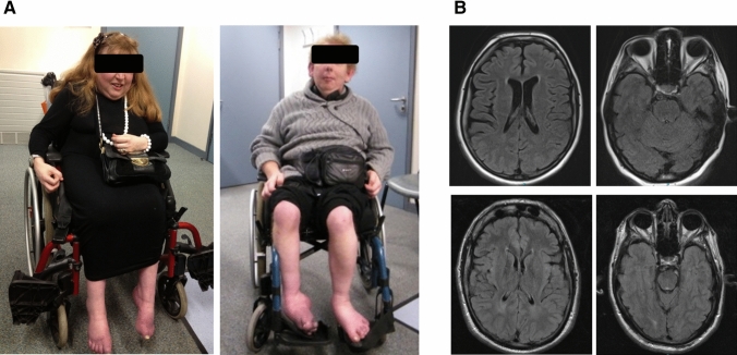
Fig. 1.
a P1 aged 40 years (left) and P2 aged 38 years. Both patients have characteristic facial features low-set ears and, and presented with distal upper and lower limb wasting and equinovarus deformity due to peripheral neuropathy and spastic paraparesis. b Axial T2 FLAIR MR images of P1 (top panels) and P2 (bottom panels). MRI brain in P1 aged 41 years showed mild global atrophy. MRI brain in P2 aged 34 demonstrated no significant abnormality apart from mild increased T2 signal in the peritrigonal white matter bilaterally

